ISU/USTUR Graduate Student Auto-Segments Case 0102 Head Phantom
George Tabatadze, M.S., a Ph.D. candidate in the Idaho State University (ISU)/USTUR Internal Dosimetry Research Group, with the help of medical physicists at the Portneuf Medical Center, Pocatello, ID, has successfully created 3-dimensional voxel images of the regions of interest in the DOE Case 0102 241Am head phantom. He used the complete set of contiguous 2-dimensional CT-scan slice (DICOM) images produced during Ms. Deanna Hasenauer’s 2006 practicum study.
Case 0102 DICOM Images
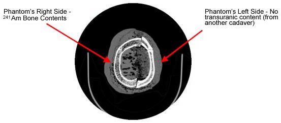
As part of her 3-month DOE Practicum study at USTUR, Ms. Hasenauer, M.S. (University of Florida) worked with Kadlec Medical Center’s Diagnostic Imaging Unit, Richland, WA to produce complete sets of contiguous, 0.625-mm CT-slice image files of the USTUR Case 0102 head, arm, torso and leg phantoms. The above diagram shows the DICOM image of a transverse slice of the head phantom. The right half of the skull consists of the actual skull bone from Case 0102 (containing 622 ± 20 Bq 241Am)1. The left half consists of unlabeled bone from a different cadaver.
Deana’s DOE practicum voxel phantom study
Segmentation, Region of Interest, and 3-D Voxel Model
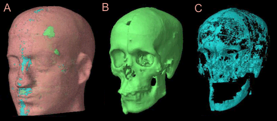
Mr. Tabatadze segmented the DICOM images of the USTUR case 0102 head phantom using Eclipse® radiotherapy planning software (Varian Medical, Palo Alto, CA). This software has a powerful automatic segmentation feature.
The three-step procedure involved:
- Defining the regions of interest as well as CT numbers for different anatomical structures
- Auto-segmenting
- Checking manually and resegmented if necessary.
Each DICOM image was segmented into the following regions of interest:
- Air Pockets
- Cortical Bone
- Bone Cavities – Marrow/Tabecular spongiosa
- Tissue – Tissue regions are subdivided into Light and Regular due to inhomogeneities that occurred when the case 102 head phantom was cast in nominal ICRU tissue equivalent plastic.
The range of CT numbers corresponding to each region of interest was replaced with a single CT number to characterize each region. Finally, all segmented DICOM images were combined into a three-dimensional ‘voxel’ phantom of the USTUR 0102 head phantom.
The segmented images were created with the great help of Dr. Steven Todd and David Theel (dosimetrist) at the Portneuf Medical Center in Pocatello, Idaho. They use the Eclipse® software to plan different radiation therapy procedures, including intensity-modulated radiation therapy (IMRT).
Reference
- U.S. Department of Energy Phantom Library: USTUR Americium-241 Bone Phantom. http://www.pnl.gov/phantom/bone/#table1-kBq. Accessed October 2008.