Intercalibration Phantoms from USTUR Cases
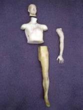
Case 0102 – A ‘Real’ 241Am Phantom
 |
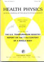 |
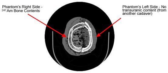
Case 0102 was the first whole body donation to the U.S. Transuranium Registry (in 1979). This case was published in 1985 – in a special issue of the Health Physics journal (Volume 49, Number 4; October 1985) dedicated to the donor. Half of this gentleman’s skeleton, encased in tissue equivalent plastic, still provides a unique anthropomorphic ‘phantom’ for calibrating whole body counting systems at DOE laboratories – and internationally.
State-of-the-Art External Counting of USTUR Donors
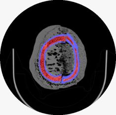
In collaboration with the Pacific Northwest National Laboratory’s (PNNL’s) ‘In Vivo’ Monitoring Program, USTUR is now using high-resolution planar germanium detectors to ‘count’ 241Am activity in various parts of donated whole bodies, before tissues and organs are sampled for radiochemical analysis. For each individual case, and also for the Case 0102 ‘real 241Am phantom,’ USTUR aims to develop a three-dimensional mathematical model, or ‘virtual phantom.’ These virtual phantoms will represent the bones of the torso, head and limbs, their radionuclide content, and the overlying thickness of soft tissue. Their availability will allow the counting efficiency of different detector types and configurations to be modeled (calculated accurately) for people of different anatomical build and body size.
Americium-241 is present as a 'contaminant' radionuclide in the majority of USTUR Registrants with accidental plutonium intakes. It emits a 59.5 keV photon which can be detected outside the body (if the activity is high enough). However, at this relatively low photon energy, the amount of self-absorption in body tissues depends rather critically on tissue thickness (body build) and the distribution of the activity within the body.
Building a ‘Virtual’ Phantom – DOE Practicum Study
 |
 |
Dr. Wes Bolch’s Advanced Laboratory for RAdiation Dosimetry Studies (ALRADS) at the University of Florida is developing a suite of skeletal dosimetric phantoms for nuclear medicine patients aged from infancy to young adulthood. These are based on computer tomographic scan (CT-Scan) images taken post mortem. The ALRADS researchers have developed mathematical methods and software codes to render a series of two-dimensional CT-Scan image files into a three-dimensional voxel model of the whole body, including the trabecular and cortical bone structure of all parts of the skeleton. Among other activities, Dr. Bolch is contributing these ‘virtual phantom’ models to the ongoing work of the International Commission on Radiological Protection’s (ICRP’s) Task Group on Internal Dosimetry (INDOS). The three-dimensional modeling techniques developed by ALRADS are directly applicable to USTUR’s case studies – of elderly donors.
The Registries are therefore especially fortunate to host one of ALRADS’ graduate students, Deanna Hasenauer, for a 3-month DOE-sponsored practicum study with the aim of initiating ‘virtual phantom’ modeling of selected USTUR whole body cases.
Presentation of Deanna’s and other current ALRADS research work
Forthcoming ALRADS presentations at the Health Physics Society’s 2006 Annual Meeting, Providence, RI, June 25-29.
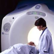
CT-Scanning – Kadlec Medical Center, Richland, WA
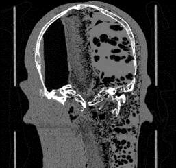
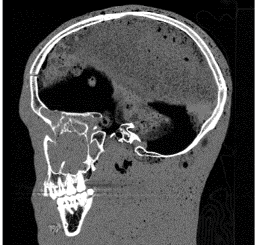
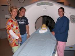
The Director and technical staff of Kadlec Medical Center’s Diagnostic Imaging Unit have volunteered to carry out the CT-scan imaging for this DOE Practicum research project. The photo shows Deanna (center) with Traci McCoy (left) and Melissa Sloan, Kadlec CT-scan specialists, preparing to run the Case #0102 skull phantom through a GE Lightspeed 16 (16 slices per rotation) scanner. See some of the initial images below – and watch this space for further developments!
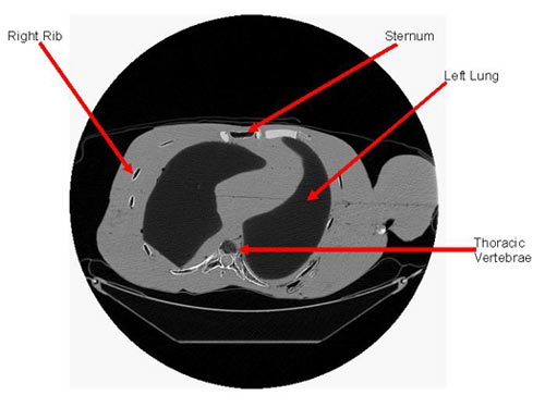
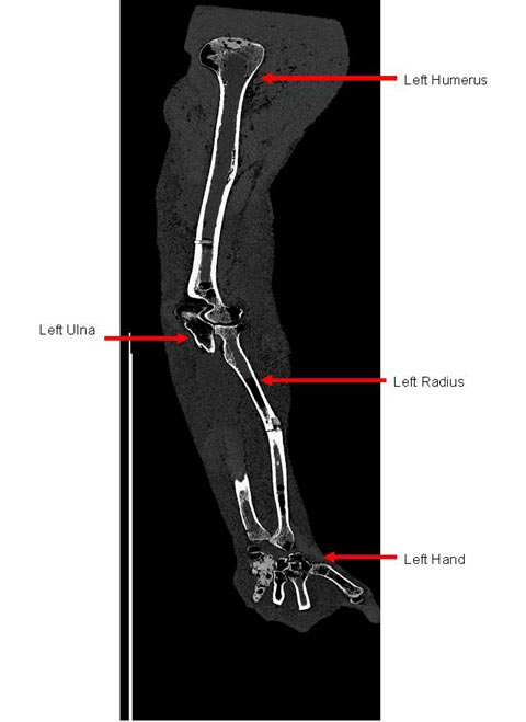
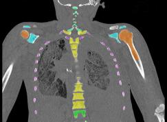
These CT slices are of the Case 0102 phantom cranium. Views were selected form the transverse, coronal, and sagital planes.