71st Annual Meeting of the Radiation Research Society, San Juan, Puerto Rico, September 21-24, 2025
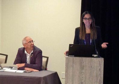
USTUR faculty gave two presentations at the 2025 Radiation Research meeting in San Juan, Puerto Rico. Sergey Tolmachev provided a case study on the biokinetics, tissue concentrations, and organ distribution of curium-244 in the human body. George Tabatadze discussed the use of the ionizing radiation quantum imaging detector (iQID) to quantify radium in human bone. Additionally, Dr. Tolmachev chaired a session on radionuclide decorporation agents and chelation therapy, and USTUR faculty were coauthors on a poster that was presented by Sara Dumit, a colleague from Los Alamos National Laboratory. Dr. Dumit’s poster summarized work to model the biokinetics of plutonium decorporation in a female USTUR Registrant.
Curium in the human body – USTUR case study
Sergey Y. Tolmachev (USTUR), George Tabatadze (USTUR), and Maia Avtandilashvili (USTUR)
Curium (Cm) is one of minor actinides presented in spent nuclear fuel. Among 19 known curium isotopes, 244Cm (T1/2 =18.1 y; Eα=5.89 MeV) accounts for more than 90% of the total curium produced in the nuclear cycle. Compared to 238Pu, 244Cm has high-power output per unit mass, and is used to manufacture radioisotope thermoelectric generators. Curium can enter the human body because of occupational incidents. Due to its high specific activity, the radiotoxicity of 244Cm predominates over the chemical toxicity. Information on curium distribution and biokinetics in the human body is limited. The United States Transuranium and Uranium Registries’ (USTUR) partial tissue donor was exposed to airborne 244Cm due to a glove-box failure. He died from hypertensive heart disease 52 years after the intake. A total of 35 soft tissue and 16 bone samples collected at autopsy were radiochemically analyzed for 244Cm. Activity concentration in systemic organs followed the pattern: skeleton>liver>kidney>muscle. The total systemic 244Cm activity was estimated to be 0.88±0.09 Bq with 91% retained in the skeleton. A total of 0.019±0.001 Bq was deposited in the respiratory tract. The activities in the respiratory tract, liver, and skeleton were used to estimate the intake and committed effective dose. The ICRP default biokinetic models, with the assumption of 83% of curium oxide, nitrate, chloride and 17% of type S material described the data well (χ2 = 1.54; p = 0.2148). Total 244Cm intake was estimated as 241 Bq and the corresponding committed effective dose was 1.71 mSv. [USTUR-0700-25A]
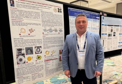
Quantifying Radium in Human Bone: A Novel Application of Digital Autoradiography
George Tabatadze (USTUR), Jessica E. Linson (University of Missouri), John D. Brockman (University of Missouri), Sergey Y. Tolmachev (USTUR)
An ionizing radiation quantum imaging detector (iQID) is employed at the United States Transuranium and Uranium Registries as a non-destructive technique for visualizing the micro-distribution of alpha-emitting radionuclides in human tissues. This study examines the potential of iQID imaging not only for spatial mapping but also for quantifying activity concentrations of 226Ra in bone samples. Two plastic-embedded bone sections, one from the middle shaft of the left femur and one from the body of the seventh thoracic vertebra, were selected from a historical case of a female radium dial painter. She had worked as a dial painter for six years, and her 226Ra intake was subsequently estimated at 58.9 MBq. She passed away at the age of 24 from diphtheria and nasopharyngeal bronchopneumonia. Cortical and trabecular bone regions were segmented, and a computational model was developed to simulate sample-specific geometric efficiencies, enabling quantitative activity concentration estimates based on alpha particle emissions detected by the iQID. The same bone sections were acid-digested and analyzed by inductively coupled plasma mass spectrometry. 226Ra activity concentrations were measured at 80.8 ± 9.2 Bq/g for the femur and 63.7 ± 17.0 Bq/g for the vertebra, providing a basis for assessing the reliability of the imaging-based estimates. This study highlights the potential of an iQID as a non-destructive technique capable of both resolving spatial heterogeneity and supporting quantitative assessments of alpha-emitting radionuclides. Such capabilities offer promising applications in internal dosimetry and retrospective dose assessment. [USTUR-0711-25A]
Modeling Plutonium Decorporation in a Female Nuclear Worker Treated with Ca-DTPA after Inhalation Intake
Sara Dumit (LANL), Maia Avtandilashvili (USTUR), Stacey L. McComish (USTUR), Guthrie Miller (retired LANL), Jasen Swanson (US Army), Sergei Y. Tolmachev (USTUR)
The present work models plutonium (Pu) biokinetics in a female former nuclear worker. Her bioassay measurements are available at the US Transuranium and Uranium Registries. The worker was internally exposed to a plutonium-americium mixture via acute inhalation at a nuclear weapons facility. She was medically treated with injections of 1 g Ca-DTPA on days 0, 5, and 14 after the intake. Between days 0 and 20, fecal and urine samples were collected and analyzed for 239Pu and 241Am. Subsequently, she was followed up for bioassay monitoring over 14 y, with additional post-treatment urine samples collected and analyzed for 239Pu. The uniqueness of this dataset is due to the availability of: (1) both early and long-term bioassay data from a female with plutonium intake; (2) data on chelation therapy for a female; and (3) fecal measurement results. Chelation therapy with Ca- and/or Zn-salts of DTPA is known to aid in reducing the internal radiation dose by enhancing the excretion of plutonium and americium from the body. Such enhancement affects plutonium biokinetics in the human body, posing a challenge to the internal dose assessment. The current radiation dose assessment practice is to exclude the data affected by Ca-DTPA from the analysis. The present analysis is the first to explicitly model the chelation-affected bioassay data in a female by using a newly developed chelation model. Thus, the bioassay data collected during and after the Ca-DTPA administrations were used for biokinetic modeling and dose assessment. The Markov Chain Monte Carlo method was used to investigate model parameter uncertainty, based on the bioassay data and assumed prior probability distributions. A χ2/nData (number of data points) ≈ 1 was observed in this study, which indicates self-consistency of the data with the model. Results of this study show that the worker’s 239Pu intake was 12 Bq, with a committed effective dose to the whole-body of 1.2 mSv and a committed equivalent dose to the bone surfaces, liver, and lungs of 37.8, 9.1, and 0.8 mSv, respectively. This study also discusses the worker’s dose reduction due to chelation treatment. [USTUR-0710-25A]